Aggregate 109+ omental caking radiology
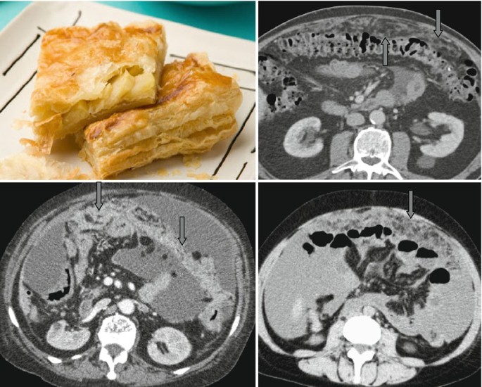
Details images of omental caking radiology by website glassplus.com.vn compilation. Gastric adenocarcinoma with omental cake | Radiology Case | Radiopaedia.org. Peritoneal Carcinomatosis and Mimicking on CT Scan Findings. Cureus | Omental Infarction: The Great Impersonator | Article. Cytoreductive Surgery and HIPEC Patient Testimonials | Stony Brook Medicine. Edwin P. Savage, MS4, 2023 RADY 401 Case Presentation: Peritoneal Carcinomatosis
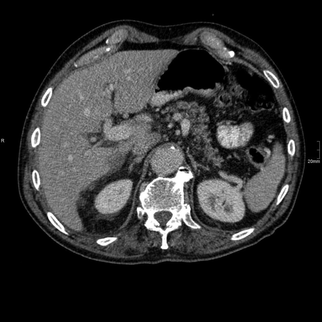 Lower gastrointestinal bleeding as a presentation of miliary tuberculosis | Gastroenterología y Hepatología – #1
 Radiology Quiz 84459 | Radiopaedia.org – #2

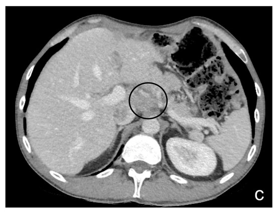 The Radiology Assistant : Acute Abdomen in Gynaecology – Ultrasound – #4
 Case 27-2016 — A 71-Year-Old Woman with Müllerian Carcinoma, Fever, Fatigue, and Myalgias | NEJM – #5
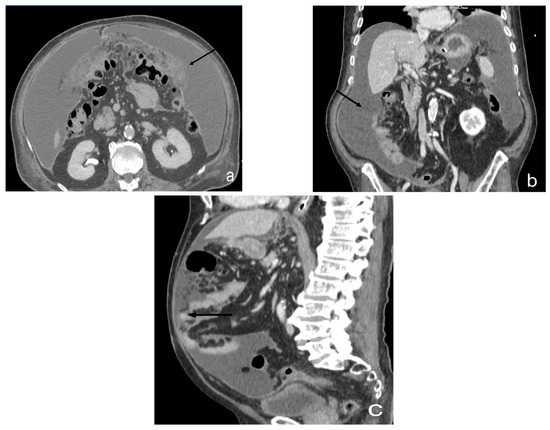 CT and MR imaging of the greater omentum: Pictorial essay – #6
 Computed tomography diagnosis of omental infarction presenting as an acute abdomen – ScienceDirect – #7
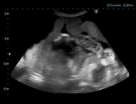 Abdominal Tuberculosis – Imaging Findings – #8

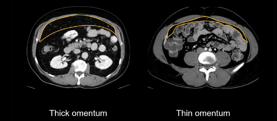
- normal omentum ct
- omental caking gross
- omental caking omental cake ultrasound
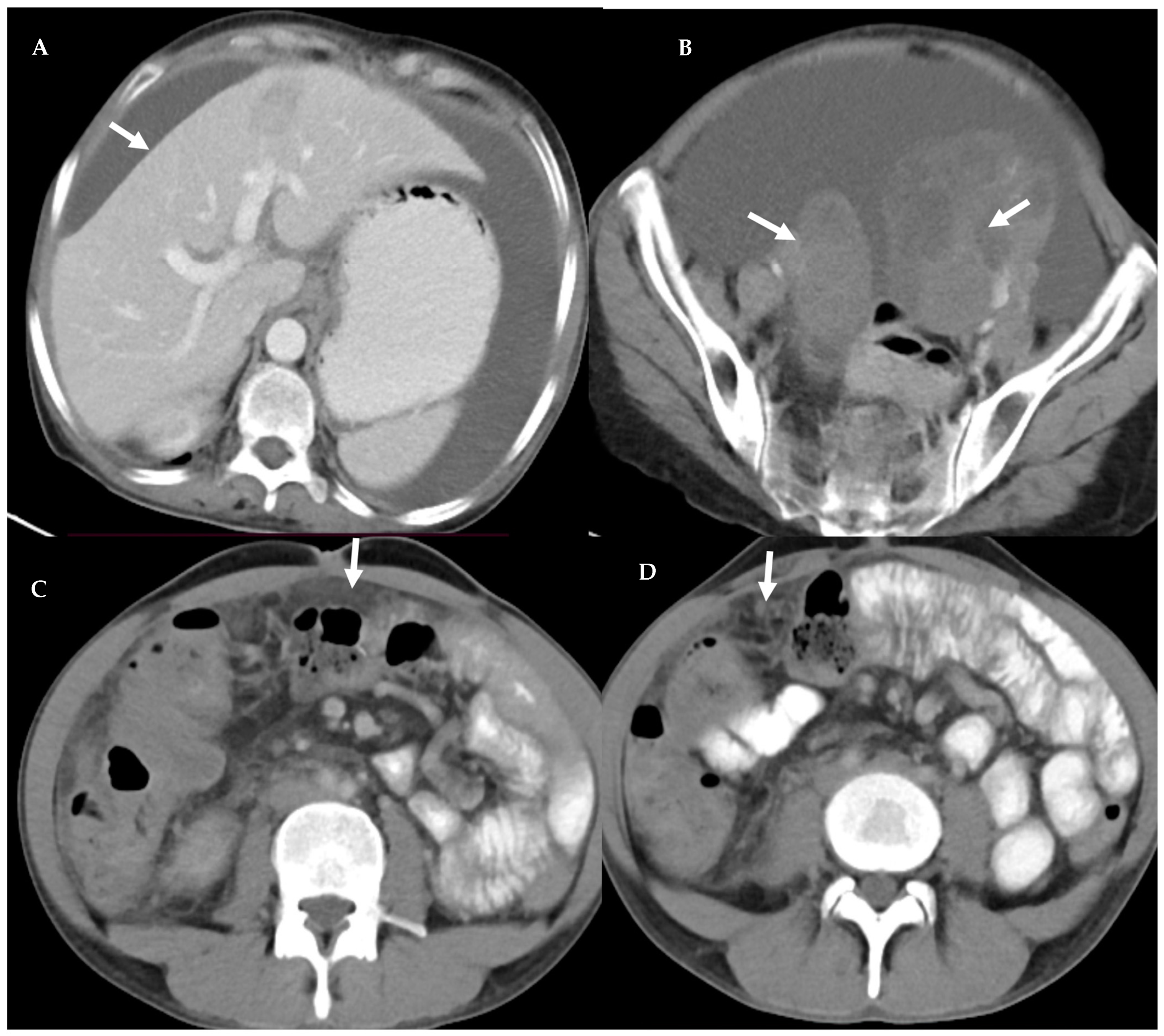 Epiploic Appendagitis: An Entity Frequently Unknown to Clinicians—Diagnostic Imaging, Pitfalls, and Look-Alikes | AJR – #10
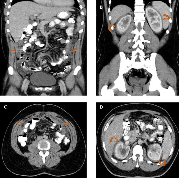 Abdominal CT: peritoneal cavity • LITFL • Radiology Library – #11
 Türk Kolon ve Rektum Hastalıkları Dergisi – #12
 MIR Teaching file case pt141 – #13
 Peritoneal carcinomatosis – Body MR Case Studies – CTisus CT Scanning – #14
 Tuberculous peritonitis mimicking carcinomatosis peritonei: CT findings and histopathologic correlation – ScienceDirect – #15
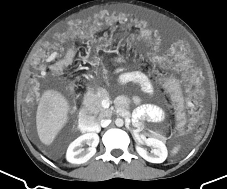 Malignant Peritoneal Mesothelioma: A Rare Cause of Ascites – Abdisamad M. Ibrahim, Mohammad Al-Akchar, Zainab Obaidi, Hamid Al-Johany, 2018 – #16
- omental caking peritoneal carcinomatosis
- stage 4 peritoneal carcinomatosis
- omental caking pathoma
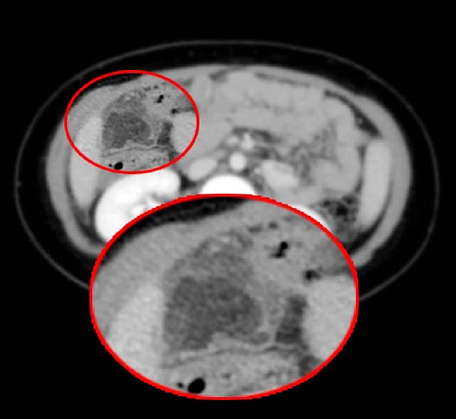 Ultrasound and computed tomography in the evaluation of mesenteric lesions: A pictorial review | SA Journal of Radiology – #17
 A rare form of presentation of non-Hodgkin lymphoma | Eurorad – #18
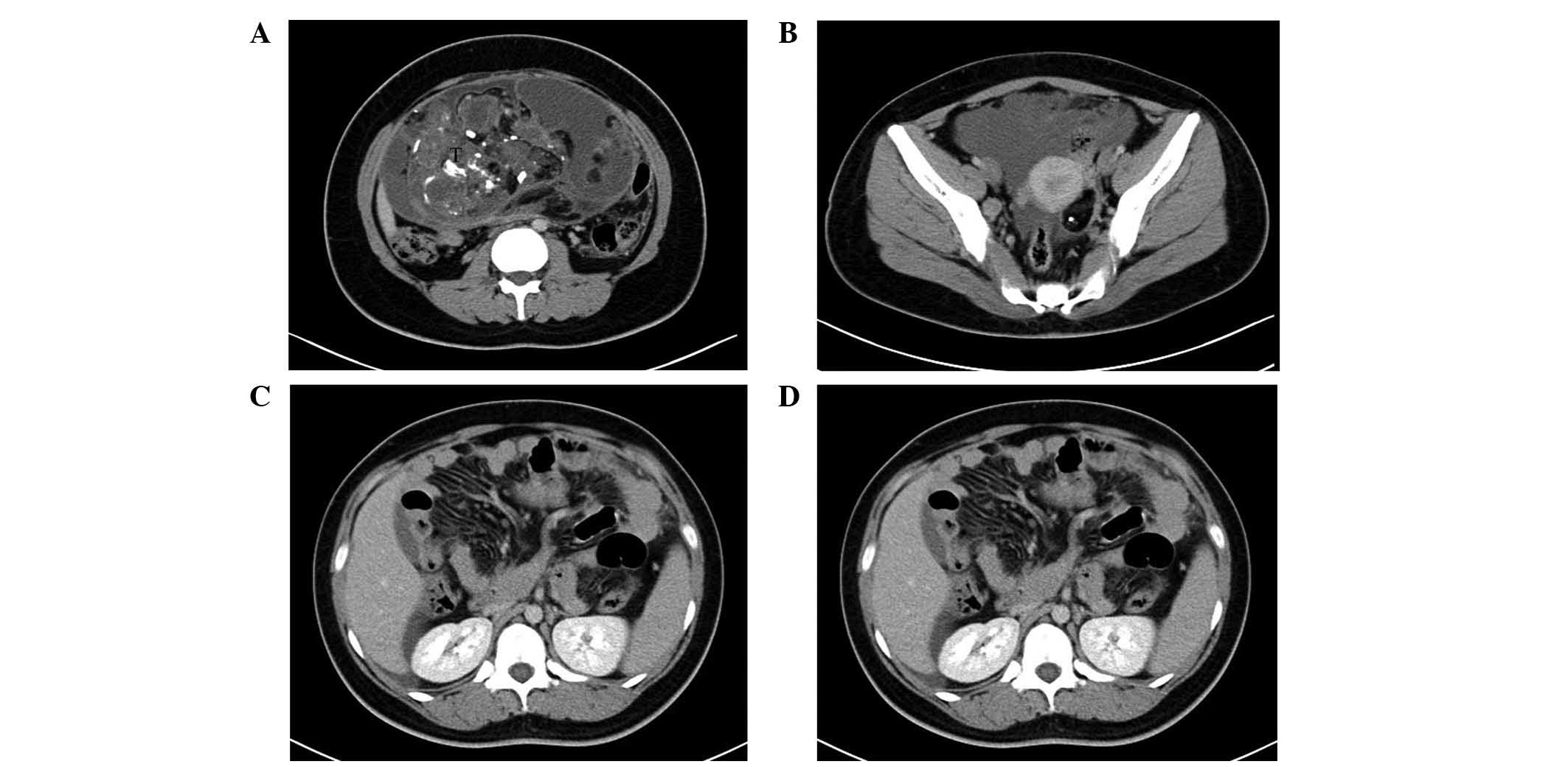
 Small fallopian tube carcinoma with extensive upper abdominal dissemination: a case report | Journal of Medical Case Reports | Full Text – #19
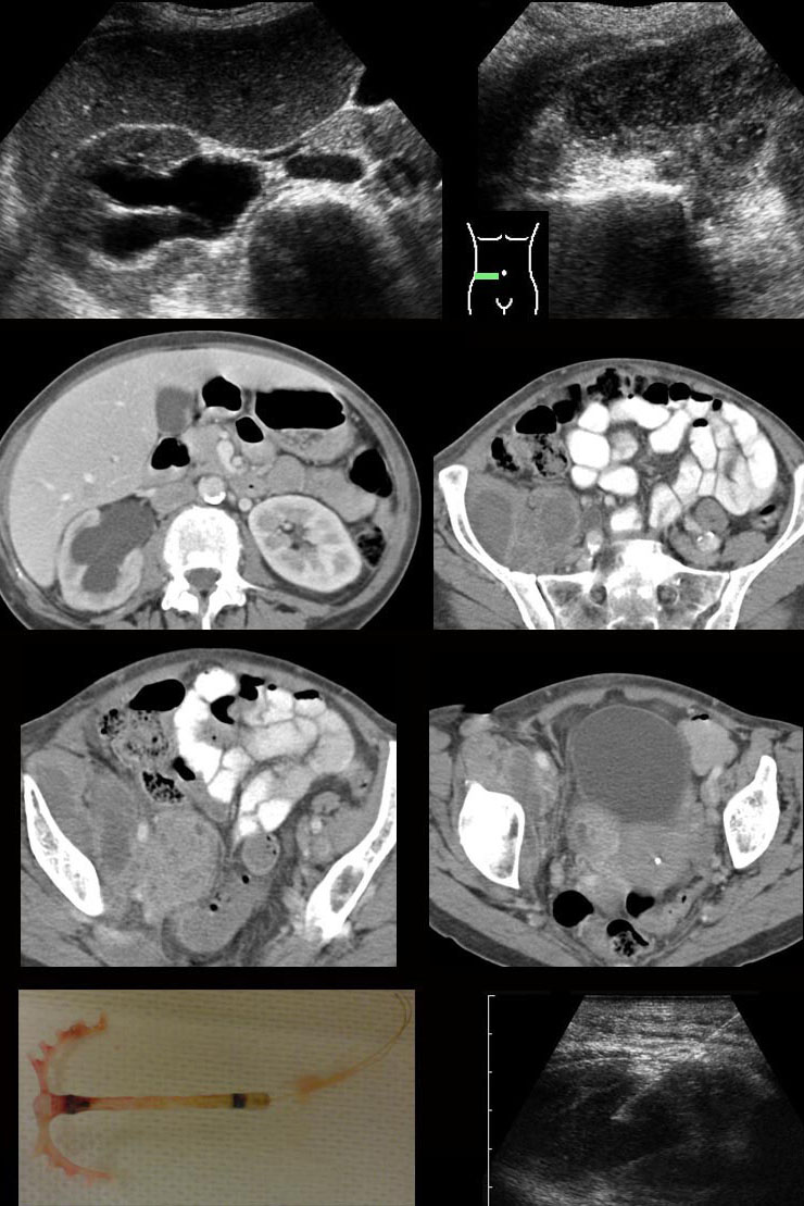 Making the most of bowel imaging with ultrasound: what Radiologist that scans often may need to know | Semantic Scholar – #20
 ACG 2023 Annual Meeting Posters – #21
 Omental Imaging Characteristics: A omental thickening, B single omental… | Download Scientific Diagram – #22
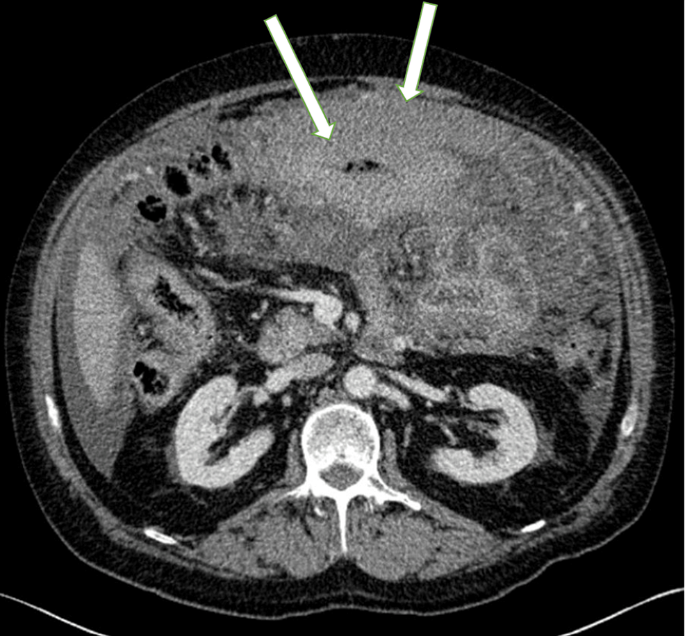 CT Scan showing omental thickening with ascitis. | Download Scientific Diagram – #23
 PDF) Omental Cake: A Radiological Diagnostic Sign – #24
 Frontiers | Case Report: Simultaneous Hyperprogression and Fulminant Myocarditis in a Patient With Advanced Melanoma Following Treatment With Immune Checkpoint Inhibitor Therapy – #25
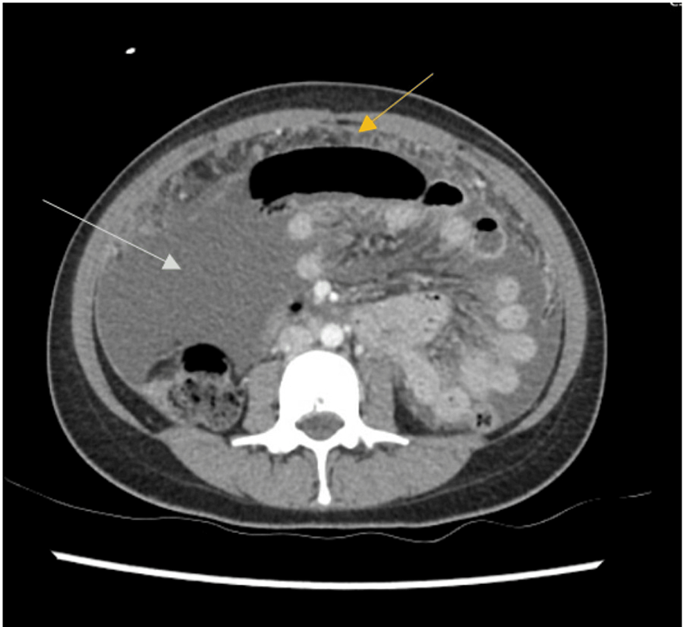 Curative Therapy for a 25 Year‐Old Woman with an Adnexal Mass and Omental Caking – SHM Abstracts | Society of Hospital Medicine – #26
 Role of Computed Tomography in the Evaluation of Peritoneal Carcinomatosis – Journal of the Belgian Society of Radiology – #27
 CT differentiation of diffuse malignant peritoneal mesothelioma and peritoneal carcinomatosis – Liang – 2016 – Journal of Gastroenterology and Hepatology – Wiley Online Library – #28
- peritoneal carcinomatosis radiology
- omental cake ultrasound
- omental caking vs normal
 Omental cakes: unusual aetiologies and CT appearances | Insights into Imaging | Full Text – #29
 Pictorial Medicine | Page 4 | HKMJ – #30
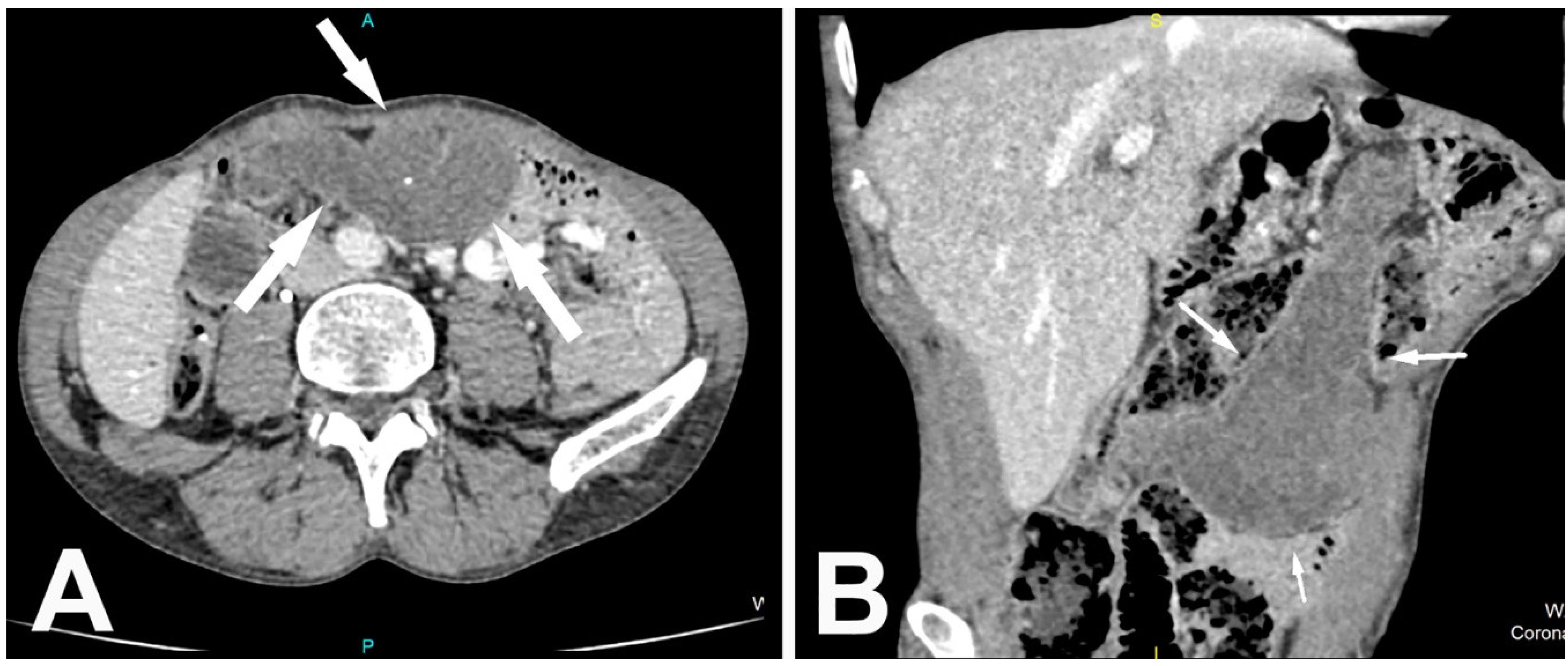 ARFI elastography of the omentum: feasibility and diagnostic performance in differentiating benign from malignant omental masses – #31
 An unusual cause of “omental cake” in CT: peritoneal tuberculosis | Eurorad – #32
 Peritoneal Sarcomatosis – radRounds Radiology Network – #33
 Peritoneal lymphomatosis: CT and PET/CT findings and how to differentiate between carcinomatosis and sarcomatosis. – Abstract – Europe PMC – #34
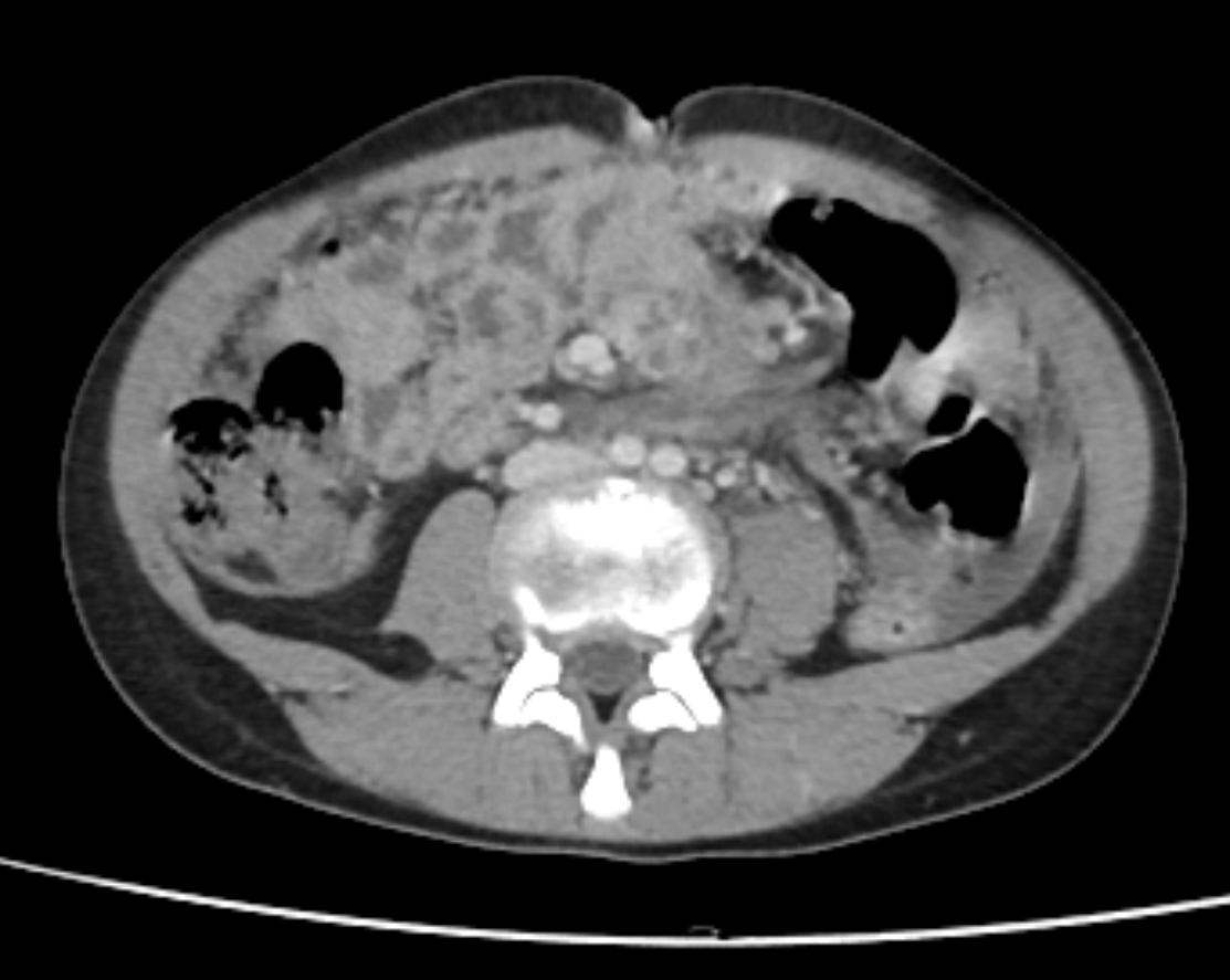 Medicina | Free Full-Text | Encapsulated Omental Necrosis as an Unexpected Postoperative Finding: A Case Report – #35
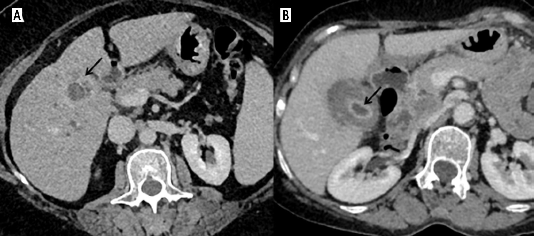 A pictorial review of the imaging findings in abdominal tuberculosis – #36
 CT scan abdomen demonstrating omental caking (arrows). | Download Scientific Diagram – #37
 Early omental cake | Radiology Case | Radiopaedia.org – #38
 Omental cake from ovarian cancer | Image | Radiopaedia.org – #39
 Colorectal peritoneal metastases: Optimal management review – #40
 Omental deposits surveillance in gynecological malignancies at first setting follow up: 18F-FDG PET/CT compared to CT – ScienceDirect – #41
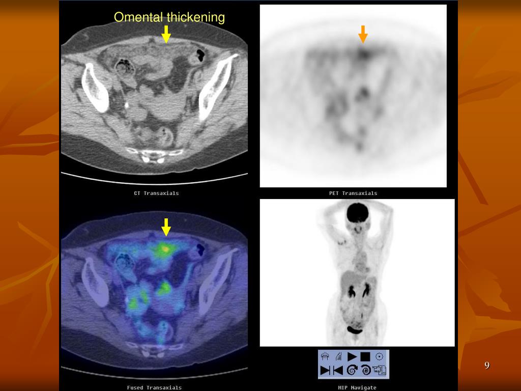 Computed tomography scan of the abdomen shows omental cake (arrows). | Download Scientific Diagram – #42
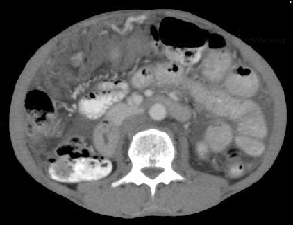 Cureus | Peritoneal Tuberculosis in Western Countries: A Rare Case With Concurrent Helminthic Infection | Article – #43
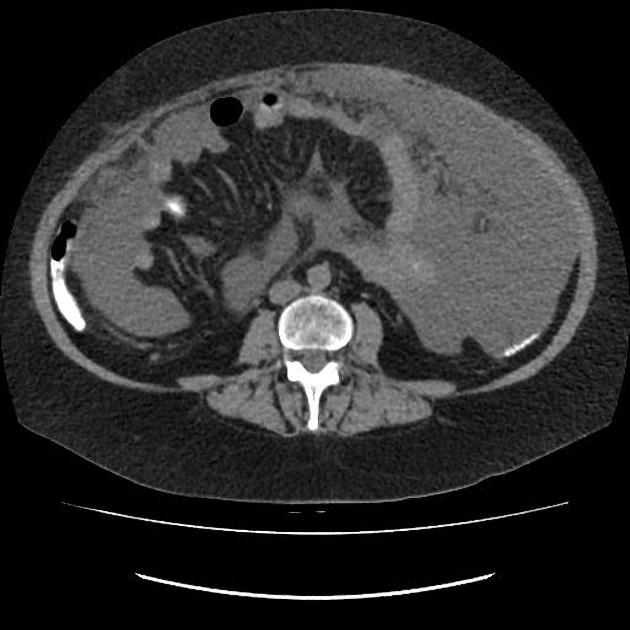 Colon cancer in a 65-year-old man. CT image demonstrates a thick… | Download Scientific Diagram – #44
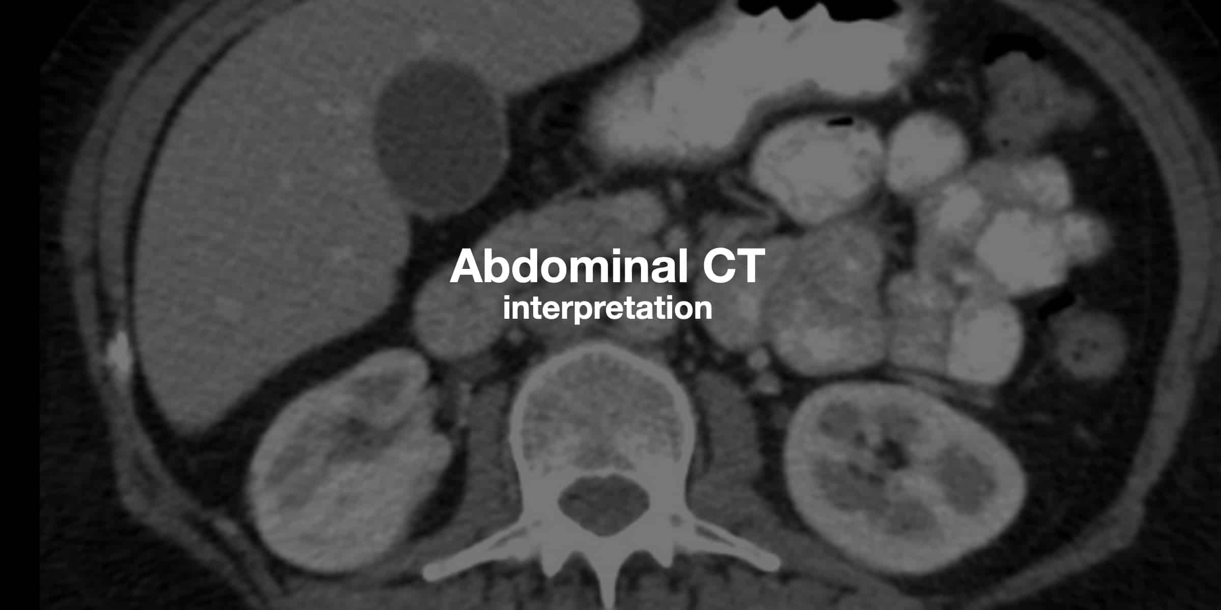 Edwin P. Savage, MS4, 2023 RADY 401 Case Presentation: Peritoneal Carcinomatosis – #45
 Omental cake – Wikipedia – #46
 Comparative Study of Ultrasound-guided Percutaneous Omental Biopsy in Cirrhotics and Noncirrhotics – #47
 Serous Surface Papillary Carcinoma of the Peritoneum: Clinical, Radiologic, and Pathologic Findings in 11 Patients | AJR – #48
 PAUSE”: a method for communicating radiological extent of peritoneal malignancy – #49
 Peritoneal carcinomatosis gastric carcinoma | Radiology Case | Radiopaedia .org – #50
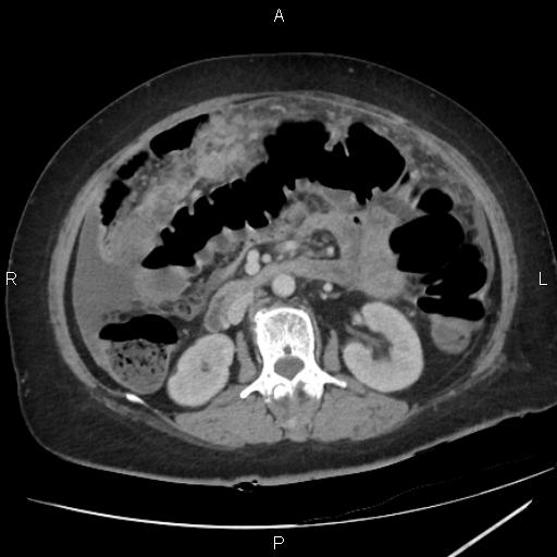 Differentiation of peritoneal tuberculosis from peritoneal carcinomatosis by the Omental Rim sign. A new sign on contrast enhanced multidetector computed tomography – ScienceDirect – #51
 Abdominal CT scan image showing confluent nodular deposits and… | Download Scientific Diagram – #52
![PDF] OMENTAL CAKING: A RARE BUT GRAVE SIGN IN PROGNOSIS OF CARCINOMA ENDOMETRIUM | Semantic Scholar PDF] OMENTAL CAKING: A RARE BUT GRAVE SIGN IN PROGNOSIS OF CARCINOMA ENDOMETRIUM | Semantic Scholar](https://upload.wikimedia.org/wikipedia/commons/9/95/CT_of_peritoneal_carcinomatosis_with_omental_smudge.jpg) PDF] OMENTAL CAKING: A RARE BUT GRAVE SIGN IN PROGNOSIS OF CARCINOMA ENDOMETRIUM | Semantic Scholar – #53
PDF] OMENTAL CAKING: A RARE BUT GRAVE SIGN IN PROGNOSIS OF CARCINOMA ENDOMETRIUM | Semantic Scholar – #53
- omental caking causes
- omental caking surgery
- omental caking picture
 Omental cake from metastatic lung carcinoma – Radiology Cases – #54
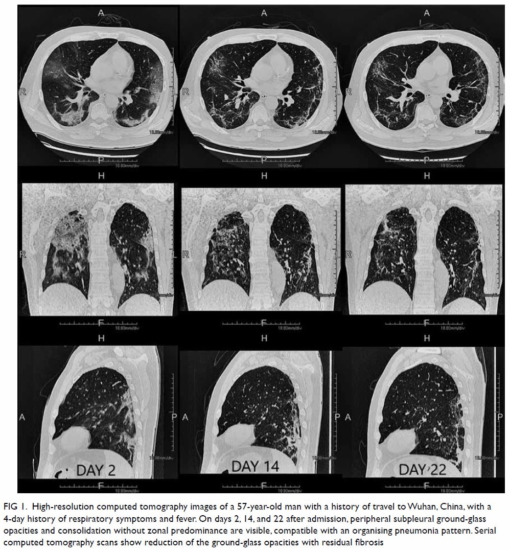 Omental cake. Enhanced transversal (a) and coronal (b) CT images of a… | Download Scientific Diagram – #55
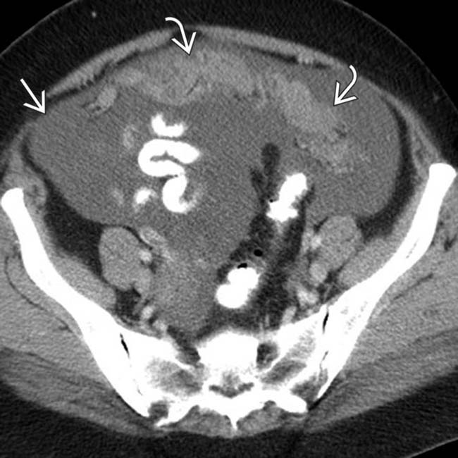 Clinical and Radiological Parameters to Discriminate Tuberculous Peritonitis and Peritoneal Carcinomatosis – #56
 Ct-scan: Burkitt´s lymphoma in the small intestine – DocCheck – #57
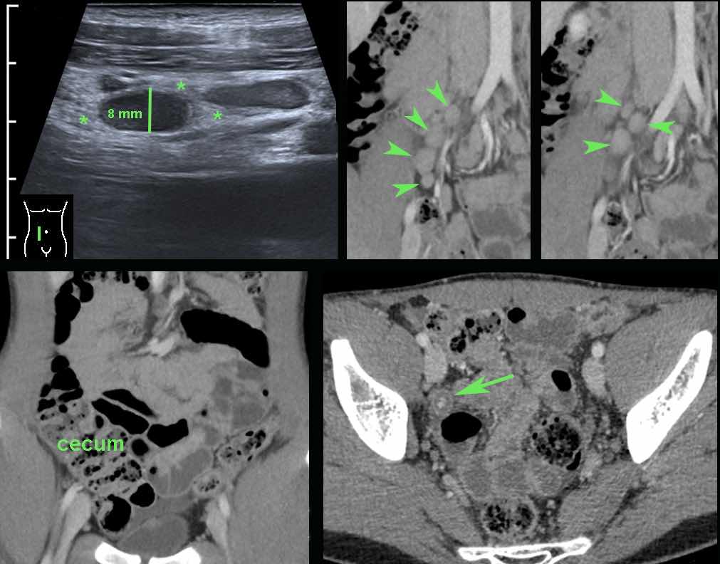 Peritoneal Carcinomatosis and Mimicking on CT Scan Findings – #58
 Greater and Lesser Omenta: Normal Anatomy and Pathologic Processes | RadioGraphics – #59
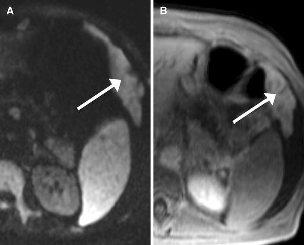
 Differentiation of Malignant Omental Caking from Benign Omental Thickening using MRI | Abdominal Radiology – #60
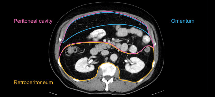 Abdomen and retroperitoneum | 1.7 Peritoneum mesentery and omentum : Case 1.7.5 Mesenteric and peritoneal metastases | Ultrasound Cases – #61
 Omental tumor. (a-e) Ultrasound, magnetic resonance with… | Download Scientific Diagram – #62
 Genevieve Crane, MD, PhD on X: “Bilateral ovarian masses, ascites, omental caking-follicular lymphoma mimicking carcinomatosis #radiology #pathology https://t.co/eU1qYEQa3O” / X – #63
 Peritoneal Metastases | Radiology Key – #64
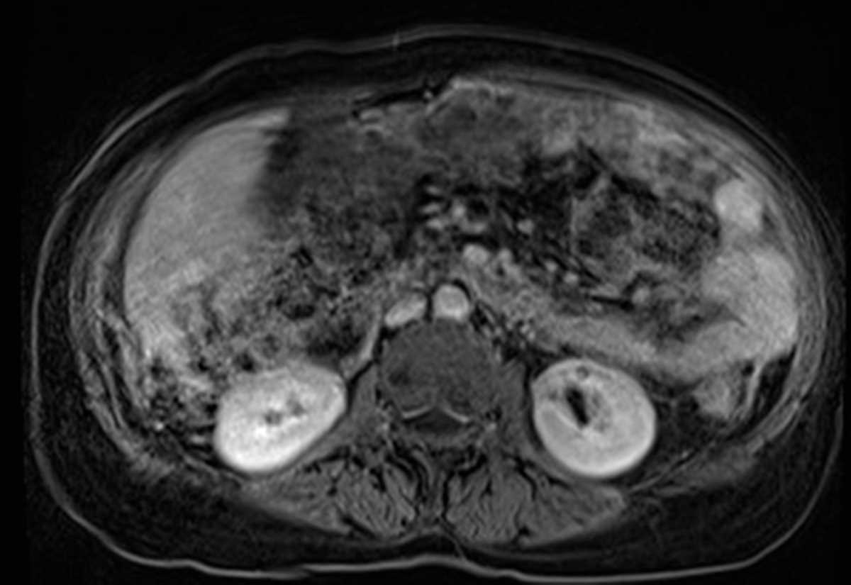 Diagnostic value of multidetector computed tomography in differentiation of benign and malignant omental lesions – #65
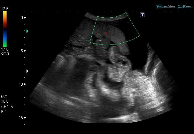 a) Drawing shows the pathways along which ovarian cancer cells with… | Download Scientific Diagram – #66
 Radiomics analysis based on CT’s greater omental caking for predicting pathological grading of pseudomyxoma peritonei | Scientific Reports – #67
 Case of the Month: Department of Radiology: Feinberg School of Medicine – #68
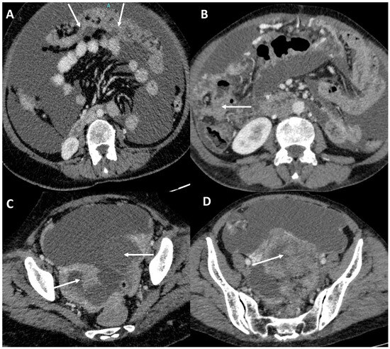 Welcome to LearningRadiology – radiology, radiologic, imaging, sign, signs, list, radiographic, xray, x-ray, gi, gu, chest, thoracic, teaching, website, web, site, best, image, student, medical, jpg, jpeg – #69
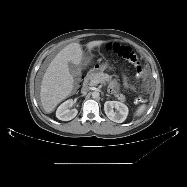 You can’t have your cake and eat it too | Gut – #70
 CT scan showing omental caking and several peritoneal implants… | Download Scientific Diagram – #71
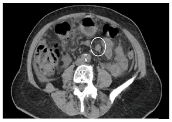 Diagnostics | Free Full-Text | Peritoneal Carcinosis: What the Radiologist Needs to Know – #72
 Peritoneal carcinomatosis: radiologic diagnosis | Advances in the Management of Peritoneal Carcinomatosis – #73
 A Case of Erdheim-Chester Disease with Omental Caking Initially Mistaken for a Malignancy | IJ Radiology | Full Text – #74
 Differentiation of peritoneal tuberculosis from peritoneal carcinomatosis by the Omental Rim sign. A new sign on contrast enhanc – #75
- omentum cancer images
- omental thickening with ascites
- ovarian cancer omental caking
 Mukesh Harisinghani on X: “Not all omental caking is due to metastases or infection; this was lymphoma https://t.co/25wwjcFVOw” / X – #76
 Testicular mesothelioma in a 53-year-old man. CT image shows an omental… | Download Scientific Diagram – #77
 Multipanel imaging of omental cake in plain radiograph, | Open-i – #78
 SciELO – Brasil – The many faces of pseudomyxoma peritonei: a radiological review based on 30 cases The many faces of pseudomyxoma peritonei: a radiological review based on 30 cases – #79
 Radiologic sign – Wikipedia – #80
 Liver Atlas: Case 29: Cholangiocarcinoma – #81
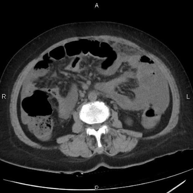 Omental cake | Radiology Case | Radiopaedia.org – #82
 Omental Infarct | Radiology Key – #83
 Omental infarction causes, symptoms, diagnosis, treatment & recovery – #84
 Omental cake from ovarian cancer | Radiology Case | Radiopaedia.org – #85
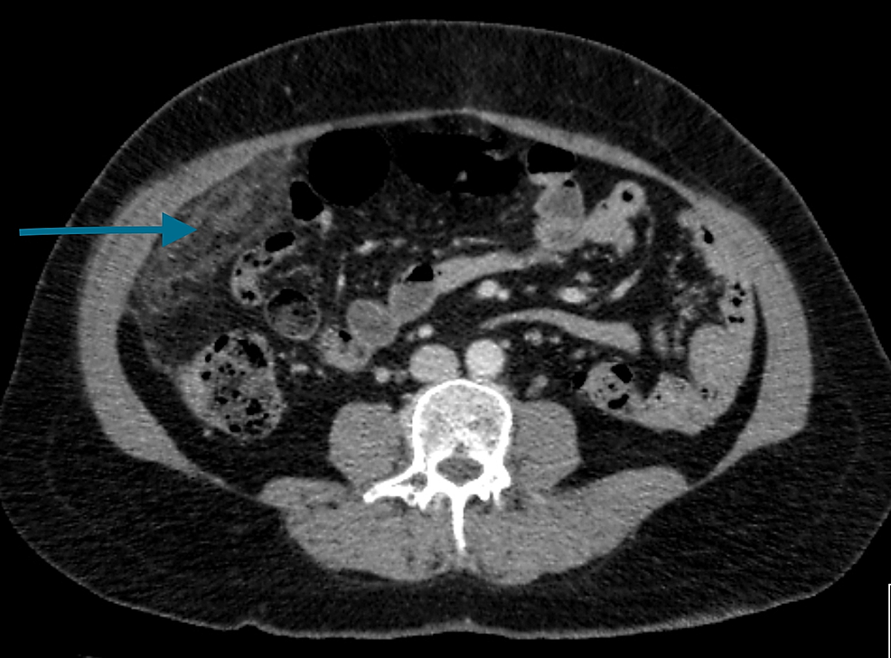 Diagnostic value of computed tomography and magnetic resonance imaging in ovarian malignant mesothelioma | BMC Medical Imaging | Full Text – #86
 Omental Cake in Peritoneal Carcinomatosis – YouTube – #87
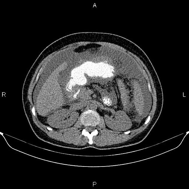 The changing face of a rare disease: lymphangioleiomyomatosis | European Respiratory Society – #88
 Diagnostics | Free Full-Text | Clinical and Radiological Parameters to Discriminate Tuberculous Peritonitis and Peritoneal Carcinomatosis – #89
 Imaging patterns of wall thickening type of gallbladder cancer – #90
 Articl net on X: “#FoamRad #AbdRad: Most viewed Radiology article on https://t.co/wKRBDCaeA4 from 2/24/2019 to 3/2/2019 – Omental cakes from Insights Imaging – https://t.co/jrvY6ZT2jK For related articles on Radiology of Omental Caking – #91
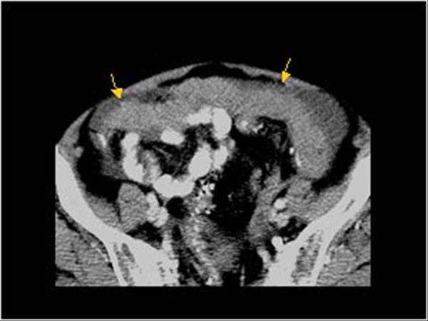 Gliomatosis peritonei with bilateral ovarian teratomas: A report of two cases – #92
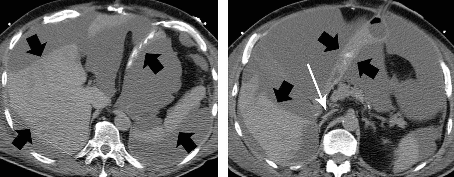
 Spectrum of computed tomography manifestations of appendiceal neoplasms: acute appendicitis and beyond – #93
 Omental Cake: A Radiological Diagnostic Sign – #94
 File:CT of peritoneal carcinomatosis with omental cake.jpg – Wikipedia – #95
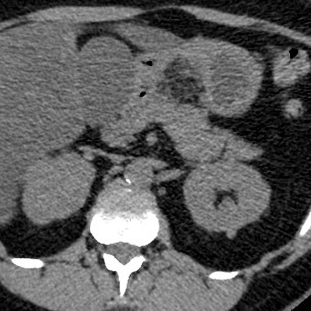
 Omental cake. : r/FOAMed911 – #96
 pelvis radiology ascites Stock Photo – Alamy – #97
 EPOS™ – #98
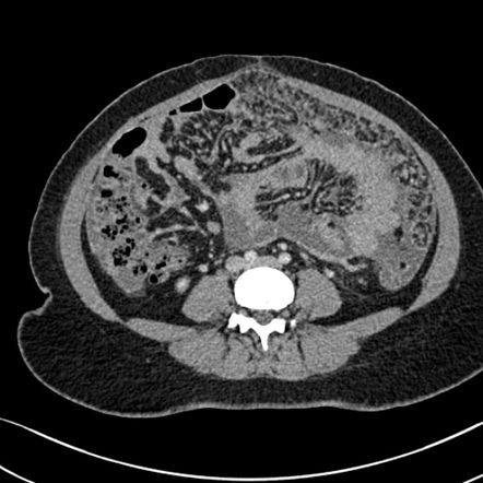 Figure 5 from Outcome of image-guided omental biopsies. | Semantic Scholar – #99
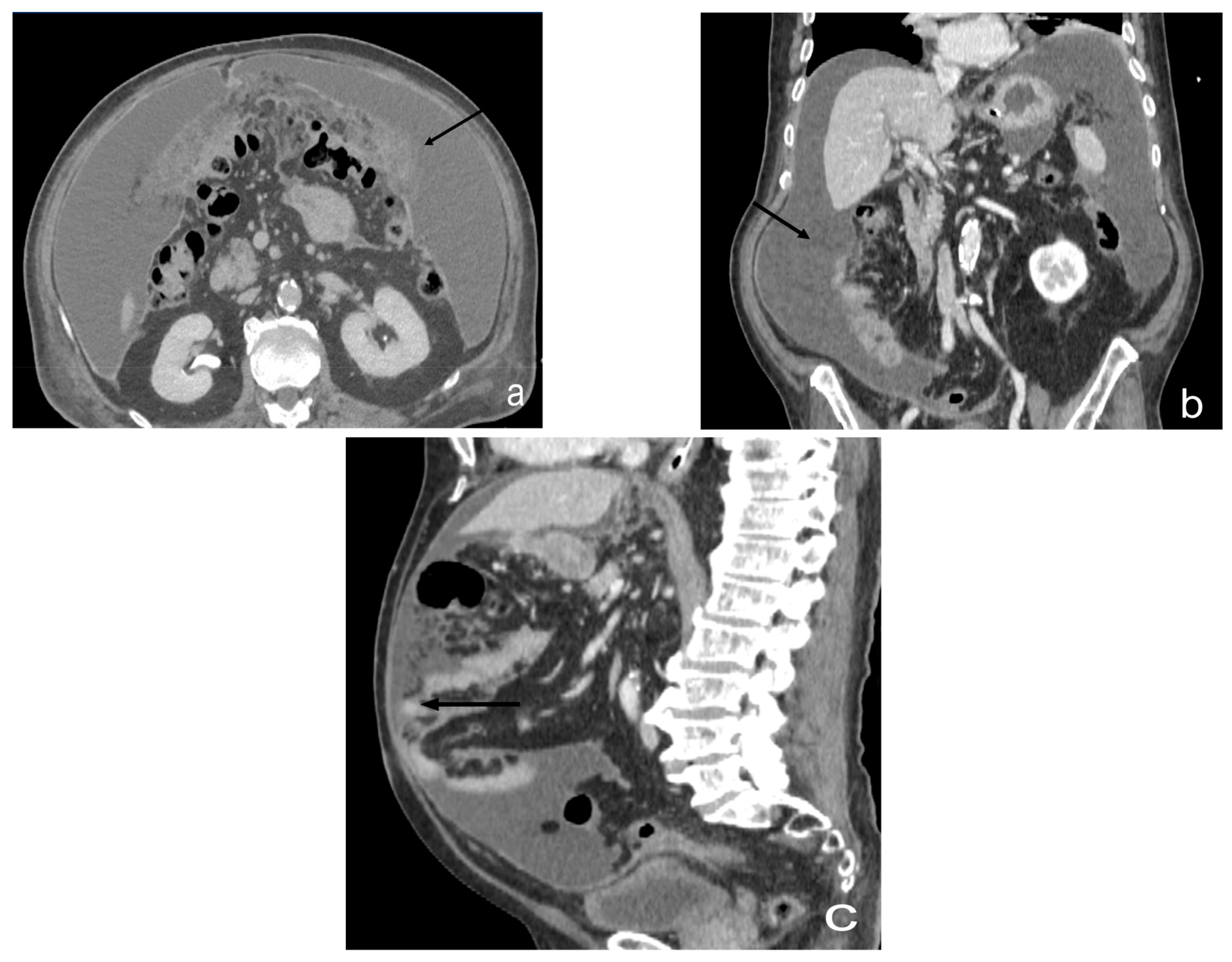 omental infarction | pacs – #100
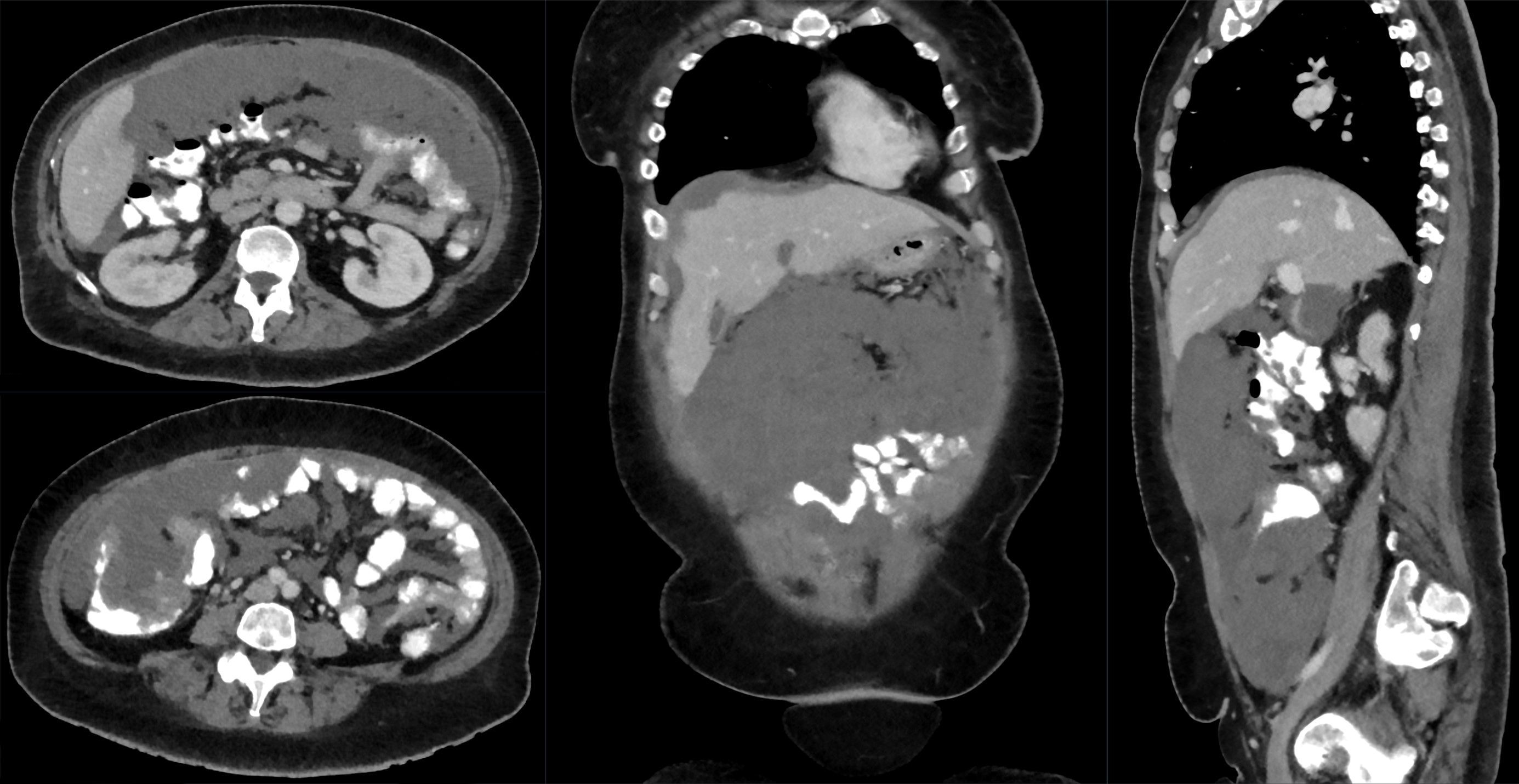 Diagnostic Approach to Benign and Malignant Calcifications in the Abdomen and Pelvis – #101
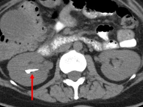 Frontiers | CT characteristics for predicting prognosis of gastric cancer with synchronous peritoneal metastasis – #102
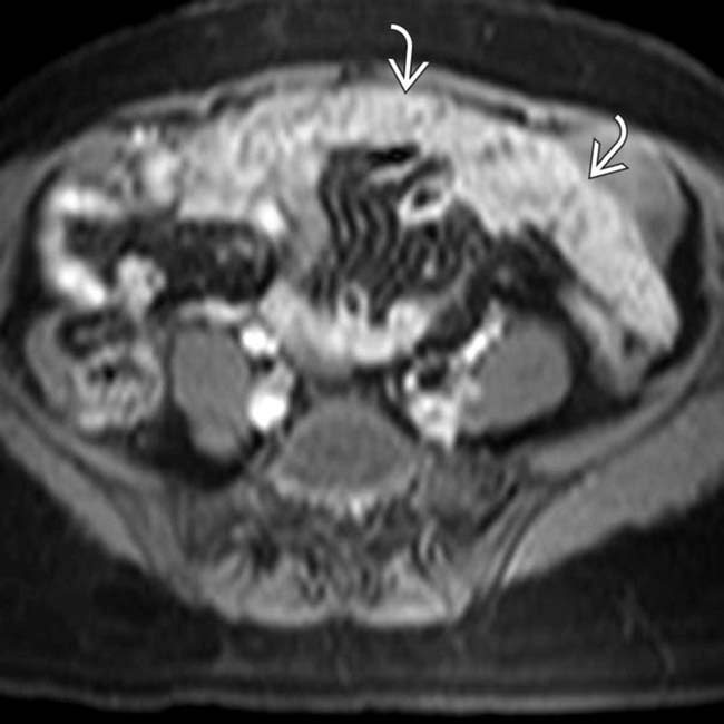 Abdomen – Radiology Cases – #103
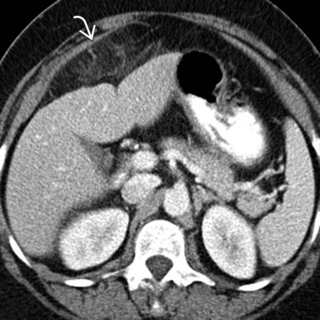
 Pseudomyxoma peritonei with omental caking. CT through the upper abdomen shows ascites with the classic scalloped liver surface (as well … | Instagram – #104
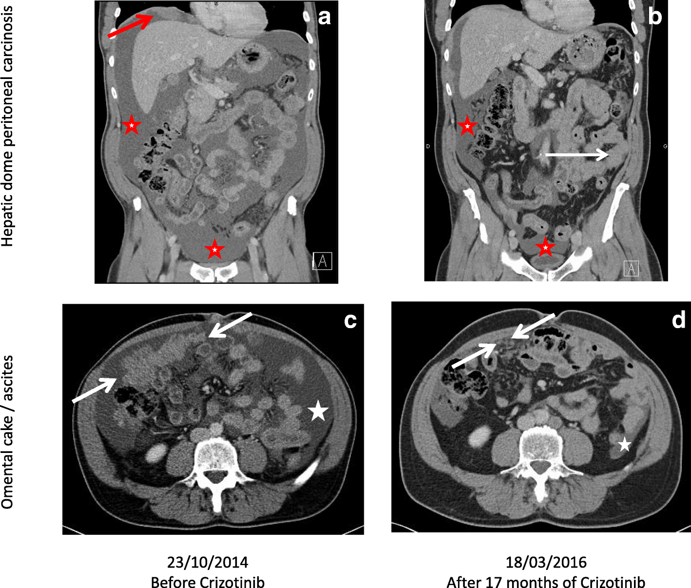
 Long-term efficacy of crizotinib in a metastatic papillary renal carcinoma with MET amplification: a case report and literature review | BMC Cancer | Full Text – #105
 PDF) CT mimics of peritoneal carcinomatosis – #106
 OMENTAL CAKE – Radiology Classroom | Facebook – #107
 Medical imaging and pathology – #108
 Unusual radiologic presentations of malignant peritoneal mesothelioma – #109
Posts: omental caking radiology
Categories: Cake
Author: glassplus.com.vn
